JOURNAL CLUB in Movement Disorders: Submandibular Gland Biopsy for Diagnosis of PD UNMC

Another interesting Journal Club with discussion on some excellent articles. Below is a brief summary of the articles with a short commentary of the discussion.

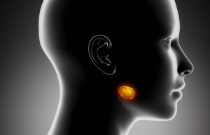
Submandibular Gland Needle Biopsy for the Diagnosis of Parkinson Disease
Neurology. 2014 Mar 11;82(10):858-64

Background: Currently Parkinson disease has no confirmatory test and diagnosis is clinical. The characteristic change in brain has been described (presence of Lewy bodies described by Fredric Lewy in 1912) however Brain biopsy is not practical in clinical settings. Braak however later described onset of changes in neurons outside the brain (called peripheral nervous system PNS) such as gut much earlier than in the brain with a spread from outside the brain to brainstem to the cortex of brain. This prompted a search for an area of the body that could be practically biopsied and is diagnostic. One such attempt earlier was biopsy of gut during colonoscopy and in another article from Japan using endoscopy.

FIG. Photomicrographs of needle core tissue from submandibular gland biopsies of subjects with PD. Section A was stained with hematoxylin and eosin. All other sections were stained with an immunohistochemical method for phosphorylated a-Syn and then counterstained with Neutral Red (see Patients and Methods). (A) Typical needle core. Arrow on left points to a region of glandular tissue whereas arrow on right points to an area of stromal connective tissue. (B) Arrow points to an immunoreactive nerve fiber within a stromal nerve fascicle. Asterisks indicate nonspecific immunoperoxidase staining of gland cell cytoplasm. (C) Immunoreactive nerve fiber adjacent to two small blood vessels (two lower asterisks). A small duct is also close by (upper asterisk is within duct lumen). (D) Several immunoreactive nerve fibers running in parallel within a stromal nerve fascicle. (E) Immunoreactive nerve fibers (arrows) within a stromal nerve fascicle. (F) Immunoreactive nerve fibers adjacent to a small duct. Asterisks indicate nonspecific immunoperoxidase staining of gland cell cytoplasm. (G) Immunoreactive nerve fiber (arrows) within a stromal nerve fascicle. (H) Immunoreactive nerve fibers (arrows) adjacent to unstained glandular epithelial cells (asterisks).

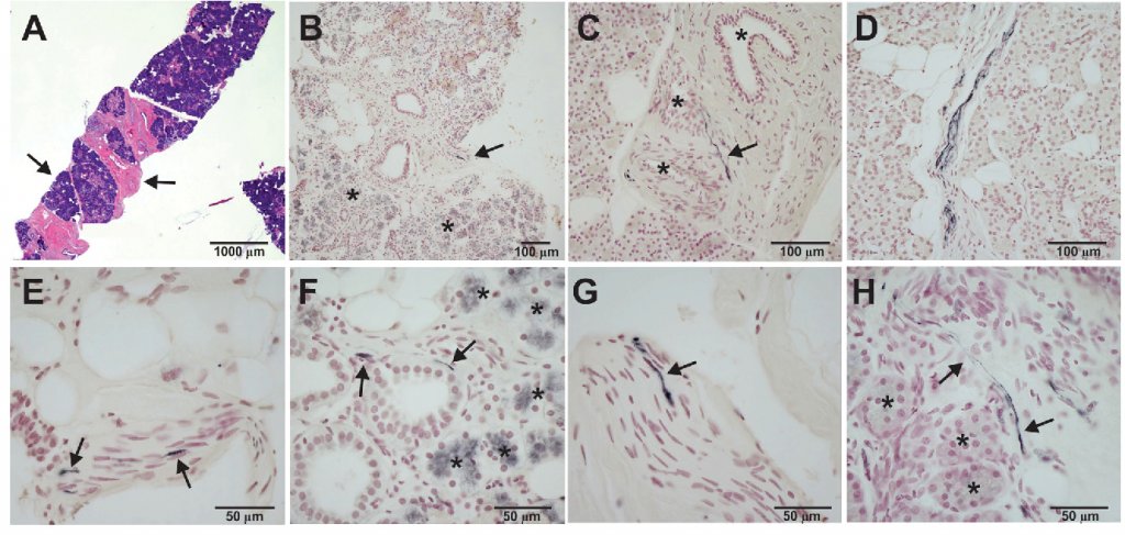
Summary: In this particular study the authors reviewed 92 autopsies with evaluation of 41 areas to look for “lewy bodies” and found all 28 patients with Parkinson disease had positive pathology in submandibular glands (inside the nerves supplying the gland). They then attempted to see if doing a needle biopsy from the submandibular gland can show the similar pathology and found a 90% success on the dead bodies. With this background they designed the current study.

15 patients (9 male and 6 female) with clinical diagnosis of Parkinson disease for more than 5 years had a submandibular gland biopsy as an outpatient procedure. The tissue obtained had submandibular gland in 12/15 patients. Of these 12 patients 9 were noted to have Parkinson disease pathology.

Discussion:
Pros – The study is remarkable in the sense that it provides an option of pathologically diagnosing the Parkinson disease that has so far not been possible. The procedure itself is a routine procedure, with minimal risk and minimally invasive. In future such an approach can also be studied for very early Parkinson disease and at risk patients.

Cons – The study has not been confirmed against a brain autopsy. The procedure did not yield submandibular gland in 3 patients and might require repeat procedures. Even after obtaining proper tissue only 9/12 were positive which may again mean requirement of repeat procedures or inherent inability of the submandibular gland biopsy to confirm diagnosis with 100% accuracy. It is also not clear if the biopsy is only positive in Parkinson disease or all neurodegenerative parkinsonisms.


I am a Movement Disorder neurologist interested in innovative medical education and use of technology in education and clinical care of my patients. My primary interest is in Parkinson Disease and am currently involved in online courses in Movement Disorders and Research with Parkinson Study Group in Neuro-protection. My hobby is biosensors and smartphone applications for diagnosis

Leave a reply:

Your email address will not be published.

Sliding Sidebar

About Me

About Me

This is a blog about various aspects of my professional side. I am a Movement Disorder Neurologist with strong focus on Parkinson's disease program development with clinical care and research, outreach and patient education. I'm an educator with a strong interest and passion in innovative methods including online learning and blended learning as they apply to clinical education or education of the medical students or more often physicians in training and practice (Continuing Medical Education). I am also using this platform to share personal achievements and sometimes general ideas which I believe are worth sharing.

I am a Movement Disorder neurologist interested in innovative medical education and use of technology in education and clinical care of my patients. My primary interest is in Parkinson Disease and am currently involved in online courses in Movement Disorders and Research with Parkinson Study Group in Neuro-protection. My hobby is biosensors and smartphone applications for diagnosis.

Subscribe to our newsletter
Sign up here to get the latest news, updates and special offers delivered directly to your inbox.
You can unsubscribe at any time